An advanced nuclear medicine imaging system that uses gamma rays to visualize organ function and detect abnormalities, supporting the diagnosis and monitoring of various diseases with high accuracy...
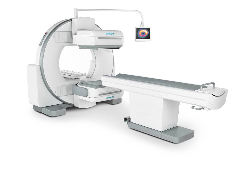
Functional Imaging Technology The gamma camera generates images by detecting the gamma rays emitted from a low-dose radiopharmaceutical administered to the patient. The Siemens ECAM Signature Series uses dedicated detectors to capture these signals and reconstruct two- or three-dimensional functional images using advanced computer processing. Imaging parameters are determined by the nuclear medicine specialist based on the organ system being evaluated, the clinical indication, and the patient's individual condition. Examination Planning All gamma camera examinations are planned by a nuclear medicine specialist following a comprehensive assessment of the patient's clinical findings and diagnostic needs. The type of radiopharmaceutical, administered dose, and imaging protocol are selected individually for each patient. Imaging Procedure During the examination, the patient is positioned beneath or adjacent to the gamma camera for the required imaging period. The system acquires imaging data non-invasively throughout the procedure. The duration of the examination varies depending on the type of nuclear medicine study being performed.



